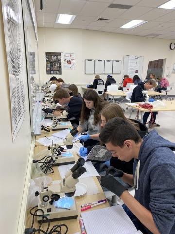


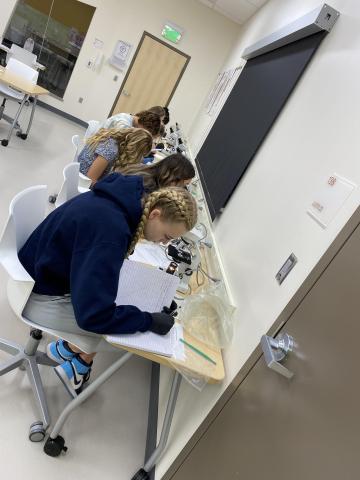
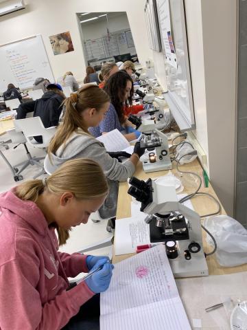
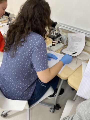
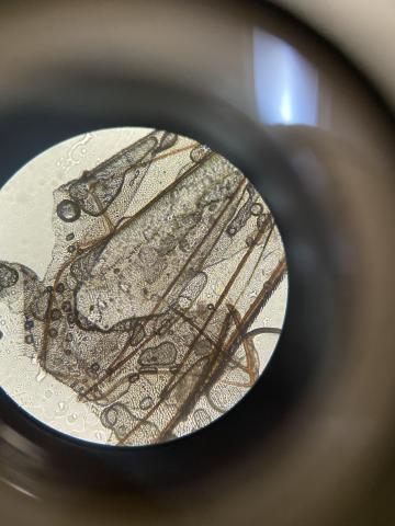
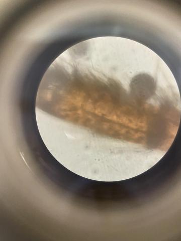
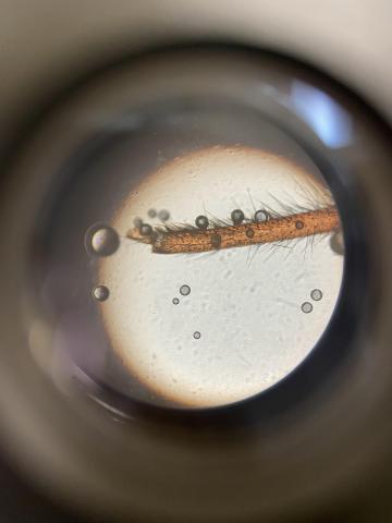
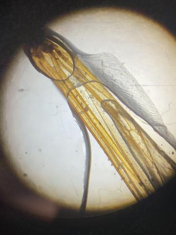
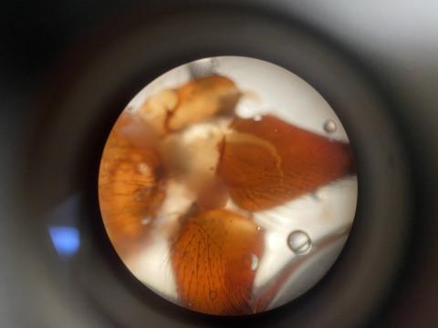
Have you ever wondered what a bug looks like magnified? Today in Medical Forensics students learned to use microscopes to see just that.
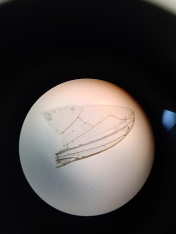
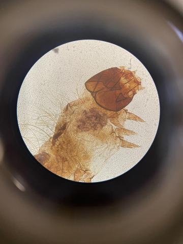
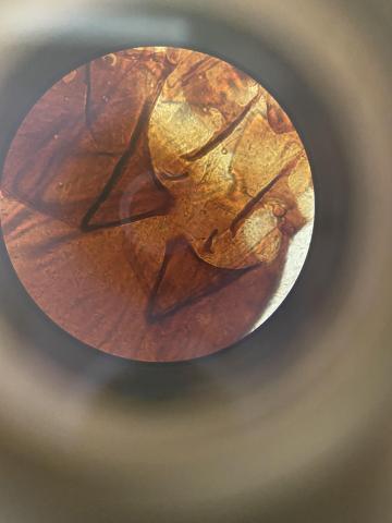
They learned to use their microscope by looking at bugs. Wings, legs, bodies, heads, and eyes.
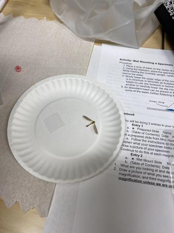
Bugs were brought in by students and then the students chose which body part they wanted to look at and prepared a slide for the microscope. The bug was viewed on three different magnifications, and students took pictures and were asked to draw what they saw.
In Medical Forensics students learn introductory principles relating to medical forensic science and crime scene investigation. This class focuses on the ability to identify, analyze, and process evidence using deductive reasoning and problem-solving. Medical forensics involves laboratory skills, microscopy, toxicology, fingerprinting, hair and fiber analysis, pathology, anthropology, entomology, criminal psychology, blood spatter analysis, and career exploration.
Once again Ms. Hartvigsen plans such great hands-on experiences for her classes. Thank you for helping our students learn in such fun, educational ways.
#ALCBeCurious #ExploreDiscoverCreate #NeboSchoolDistrict #EngageStudents
#RiseUp #NeboHero #NeboSchoolDistrict #StudentSuccess #EmpowerStudents #EngageStudents #FocusOnStudents #LoveUTpublicSchools #UtPol #UtEd #ThankATeacher #LoveTeaching